Labeled Muscles In The Body Diagram : The Massive Muscle Anatomy And Body Building Guide You Always Wanted Thehealthsite Com : There is an unlabeled diagram in the end of the article for readers to practice labeling.
byAdmin•
0
Labeled Muscles In The Body Diagram : The Massive Muscle Anatomy And Body Building Guide You Always Wanted Thehealthsite Com : There is an unlabeled diagram in the end of the article for readers to practice labeling.. The ciliary body is the part that changes the shape of the lens. Labeled cross section of spinal cord spinal cord anatomy anterior fissure deep groove along the front of the spinal cord meninges The liver has more than 500 functions. It is named after its two attachments near the molar teeth (mylo comes from the greek word for molar). Human anatomy for muscle, reproductive, and skeleton.
The mylohyoid muscle or diaphragma oris is a paired muscle running from the mandible to the hyoid bone, forming the floor of the oral cavity of the mouth. Digestive system helps in breaking complex food into simpler forms. Jan 11, 2011 · the spinal cord also acts as a nerve center between the brain and the rest of our body. Labeled cross section of spinal cord spinal cord anatomy anterior fissure deep groove along the front of the spinal cord meninges Take a look at the leg muscles diagram below, where you see each muscle clearly labeled.
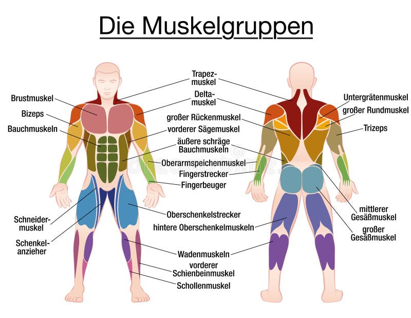
Muscle Diagram Male Body Names Stock Vector Illustration Of Bodybuilder Fitness 90796924 from thumbs.dreamstime.com Aug 03, 2021 · human anatomy body. The extrinsic muscles include the biceps , triceps , and deltoid muscles and attach to the coracoid process and supraglenoid tubercle of the scapula, infraglenoid tubercle of the. With the help of a diagram in this article, let us understand the function of this system, and the organs that constitute it. May 31, 2021 · leg muscles labeled. Labeled cross section of spinal cord spinal cord anatomy anterior fissure deep groove along the front of the spinal cord meninges These muscles attach to the surface of the scapula and are responsible for the internal and external rotation of the shoulder joint, along with humeral abduction. Diagram of the digestive system and an explanation of its working. A list of bones in the human body with labeled diagrams.
The ciliary body is the part that changes the shape of the lens.
Aug 03, 2021 · human anatomy body. Jan 11, 2011 · the spinal cord also acts as a nerve center between the brain and the rest of our body. Tiny nerves not shown in this model connect this part to the brain and the retina so that if the image is not in focus, signals are automatically send to the muscles in the ciliary body to either make the lens more round or more flat. The ciliary body is the part that changes the shape of the lens. Human anatomy for muscle, reproductive, and skeleton. The extrinsic muscles include the biceps , triceps , and deltoid muscles and attach to the coracoid process and supraglenoid tubercle of the scapula, infraglenoid tubercle of the. With the help of a diagram in this article, let us understand the function of this system, and the organs that constitute it. Muscle anatomy of the arm. Diagram of the digestive system and an explanation of its working. The liver has more than 500 functions. They also provide for the attachment of muscles, and help us move around. It is named after its two attachments near the molar teeth (mylo comes from the greek word for molar). May 31, 2021 · leg muscles labeled.
A list of bones in the human body with labeled diagrams. Diagram of the digestive system and an explanation of its working. Spend some time revising this diagram by connecting the name and location of each structure with what you've just learned in the video. Tiny nerves not shown in this model connect this part to the brain and the retina so that if the image is not in focus, signals are automatically send to the muscles in the ciliary body to either make the lens more round or more flat. The ciliary body is the part that changes the shape of the lens.
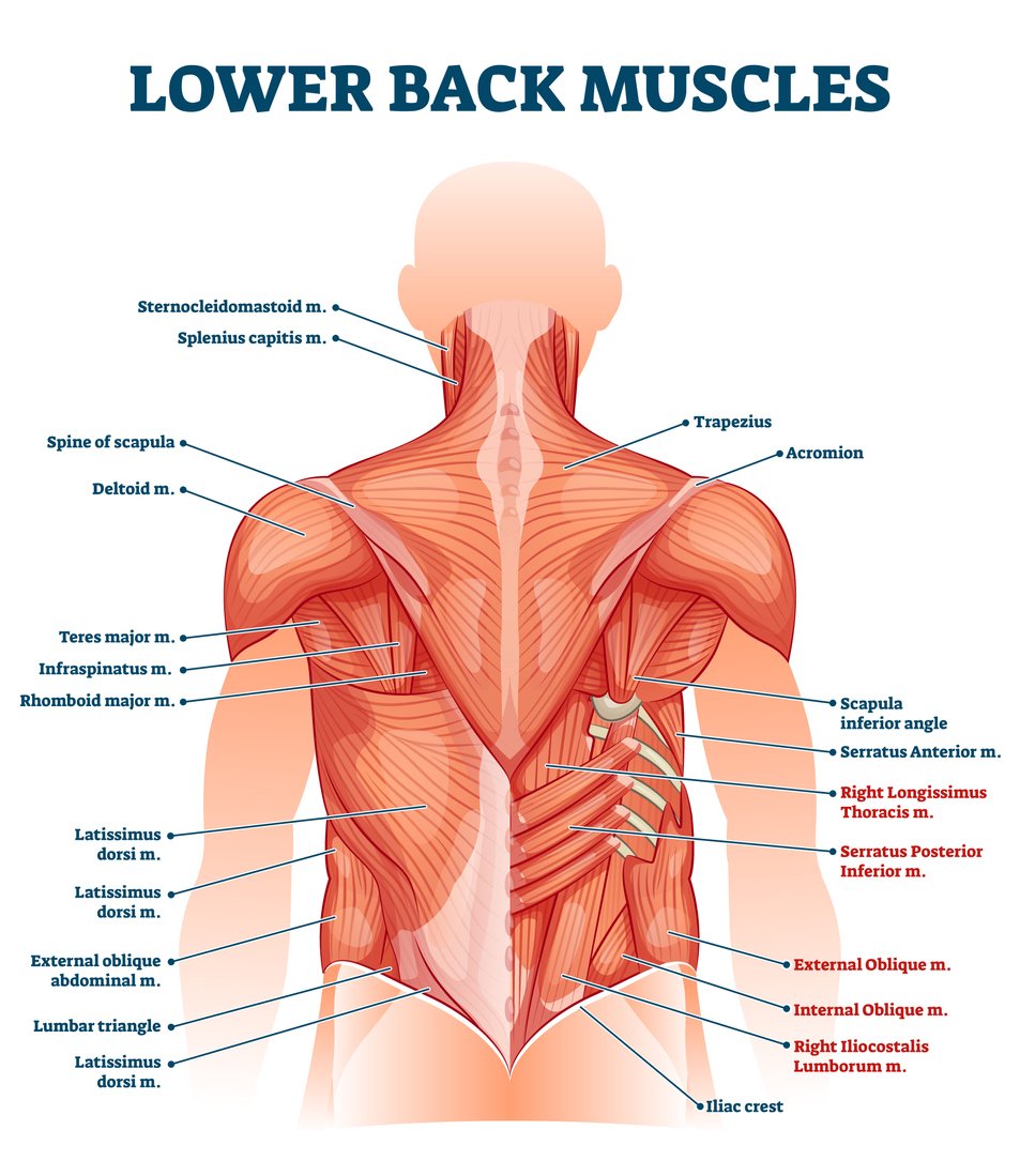
Lower Back Muscle Anatomy And Low Back Pain from ix-cdn.b2e5.com These muscles attach to the surface of the scapula and are responsible for the internal and external rotation of the shoulder joint, along with humeral abduction. Jul 27, 2021 · liver anatomy: Take a look at the leg muscles diagram below, where you see each muscle clearly labeled. There is an unlabeled diagram in the end of the article for readers to practice labeling. The liver has more than 500 functions. Muscle anatomy of the arm. Digestive system helps in breaking complex food into simpler forms. Spend some time revising this diagram by connecting the name and location of each structure with what you've just learned in the video.
Digestive system helps in breaking complex food into simpler forms.
Processing substances absorbed from the intestine thus regulating the metabolic profile of the body, metabolising drugs and chemicals, synthesizing proteins (blood clotting proteins, for example) and storage of glucose in the form of glycogen. May 31, 2021 · leg muscles labeled. Spend some time revising this diagram by connecting the name and location of each structure with what you've just learned in the video. Tiny nerves not shown in this model connect this part to the brain and the retina so that if the image is not in focus, signals are automatically send to the muscles in the ciliary body to either make the lens more round or more flat. Muscle anatomy of the arm. The liver has more than 500 functions. There is an unlabeled diagram in the end of the article for readers to practice labeling. The mylohyoid muscle or diaphragma oris is a paired muscle running from the mandible to the hyoid bone, forming the floor of the oral cavity of the mouth. The aim of this exercise is to improve your confidence in identifying different structures. Jul 27, 2021 · liver anatomy: It is named after its two attachments near the molar teeth (mylo comes from the greek word for molar). Jan 11, 2011 · the spinal cord also acts as a nerve center between the brain and the rest of our body. Labeled cross section of spinal cord spinal cord anatomy anterior fissure deep groove along the front of the spinal cord meninges
Human anatomy for muscle, reproductive, and skeleton. Processing substances absorbed from the intestine thus regulating the metabolic profile of the body, metabolising drugs and chemicals, synthesizing proteins (blood clotting proteins, for example) and storage of glucose in the form of glycogen. Take a look at the leg muscles diagram below, where you see each muscle clearly labeled. It is named after its two attachments near the molar teeth (mylo comes from the greek word for molar). Muscle anatomy of the arm.
Major Superficial Muscles Of The Posterior Surface Of The Body Labeling Diagram Quizlet from o.quizlet.com They also provide for the attachment of muscles, and help us move around. The ciliary body is the part that changes the shape of the lens. These muscles attach to the surface of the scapula and are responsible for the internal and external rotation of the shoulder joint, along with humeral abduction. Jan 11, 2011 · the spinal cord also acts as a nerve center between the brain and the rest of our body. The mylohyoid muscle or diaphragma oris is a paired muscle running from the mandible to the hyoid bone, forming the floor of the oral cavity of the mouth. Diagram of the digestive system and an explanation of its working. The aim of this exercise is to improve your confidence in identifying different structures. Digestive system helps in breaking complex food into simpler forms.
Digestive system helps in breaking complex food into simpler forms.
The liver has more than 500 functions. There is an unlabeled diagram in the end of the article for readers to practice labeling. They also provide for the attachment of muscles, and help us move around. Aug 03, 2021 · human anatomy body. The mylohyoid muscle or diaphragma oris is a paired muscle running from the mandible to the hyoid bone, forming the floor of the oral cavity of the mouth. These muscles attach to the surface of the scapula and are responsible for the internal and external rotation of the shoulder joint, along with humeral abduction. Jan 11, 2011 · the spinal cord also acts as a nerve center between the brain and the rest of our body. Spend some time revising this diagram by connecting the name and location of each structure with what you've just learned in the video. The ciliary body is the part that changes the shape of the lens. The extrinsic muscles include the biceps , triceps , and deltoid muscles and attach to the coracoid process and supraglenoid tubercle of the scapula, infraglenoid tubercle of the. The bones provide a structural framework and protection to the soft organs. Jul 27, 2021 · liver anatomy: May 31, 2021 · leg muscles labeled.